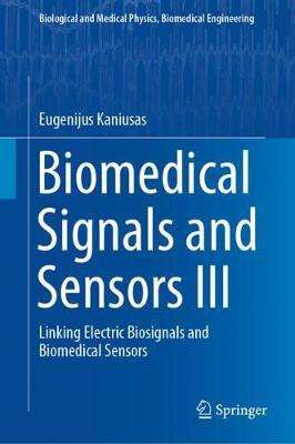
Biomedical Signals & Sensors III: Linking Electric Biosignals & Biomedical Sensors
Original price was: ₹13,874.08.₹11,099.26Current price is: ₹11,099.26.
ISBN: 9783319749167
Author/Editor: Eugenijus Kaniusas
Publisher: Springer
Year: 2019
Available on backorder
Description
As the third volume in the author? series on ?iomedical Signals and Sensors,?this book explains in a highly instructive way how electric, magnetic and electromagnetic fields propagate and interact with biological tissues. The series provides a bridge between physiological mechanisms and theranostic human engineering. The first volume focuses on the interface between physiological mechanisms and the resultant biosignals that are commonplace in clinical practice. The physiologic mechanisms determining biosignals are described from the cellular level up to the mutual coordination at the organ level. In turn, the second volume considers the genesis of acoustic and optic biosignals and the associated sensing technology from a strategic point of view. This third volume addresses the interface between electric biosignals and biomedical sensors. Electric biosignals are considered, starting with the biosignal formation path to biosignal propagation in the body and finally to the biosignal sensing path and the recording of the signal. The series also emphasizes the common features of acoustic, optic and electric biosignals, which are ostensibly entirely different in terms of their physical nature. Readers will learn how these electric, magnetic and electromagnetic fields propagate and interact with biological tissues, are influenced by inhomogeneity effects, cause neuromuscular stimulation and thermal effects, and finally pass the electrode/tissue boundary to be recorded. As such, the book helps them manage the challenges posed by the highly interdisciplinary nature of biosignals and biomedical sensors by presenting the basics of electrical engineering, physics, biology and physiology that are needed to understand the relevant phenomena.
Additional information
| Weight | 1.25 kg |
|---|
Product Properties
| Year of Publication | 2019 |
|---|---|
| Table of Contents | PREFACE ACKNOWLEDGEMENTS SYMBOLS AND ABBREVIATIONS SYMBOLS OF BIOSIGNALS 6 SENSING BY ELECTRIC BIOSIGNALS 6.1 Formation aspects 6.1.1 Permanent biosignals 6.1.2 Induced biosignals 6.1.3 Transmission of electric signals 6.1.3.1 Propagation of electric signals 6.1.3.1.1 Lossless medium 6.1.3.1.2 Lossy medium 6.1.3.2 Effects on electric signals 6.1.3.2.1 Volume effects 6.1.3.2.1.1 General issues 6.1.3.2.1.1.1 Electric and magnetic fields 6.1.3.2.1.1.2 Current density and current 6.1.3.2.1.1.3 Electric field and voltage 6.1.3.2.1.1.4 Electrical impedance 6.1.3.2.1.1.5 Simple tissue model 6.1.3.2.1.1.6 Mutual field coupling and quasi-electrostatic situation 6.1.3.2.1.2 Incident electric fields 6.1.3.2.1.2.1 Conductive phenomena 6.1.3.2.1.2.2 Polarization phenomena 6.1.3.2.1.2.3 Conductive versus polarization behaviour 6.1.3.2.1.2.4 Conductivity and polarization with relaxation and dispersion 6.1.3.2.1.2.5 Charge and current induction 6.1.3.2.1.3 Incident magnetic fields 6.1.3.2.1.4 Incident electromagnetic fields 6.1.3.2.2 Inhomogeneity effects 6.1.3.2.2.1 Boundary conditions 6.1.3.2.2.1.1 Conductive phenomena 6.1.3.2.2.1.2 Displacement phenomena 6.1.3.2.2.1.3 Conductive and displacement phenomena 6.1.3.2.2.1.4 Inhomogeneous structures and varying frequency 6.1.3.2.2.2 Diffraction 6.1.3.2.2.3 Reflection and refraction 6.1.3.2.3 Volume and inhomogeneity effects - a quantitative approach 6.1.3.2.3.1 Incindent electric field 6.1.3.2.3.1 Incident contact current 6.1.3.2.3.1 Incident magnetic field 6.1.3.2.4 Physiological effects 6.1.3.2.4.1 Stimulation effects 6.1.3.2.4.1.1 Current density versus electric field 6.1.3.2.4.1.2 Charge transfer during stimulation 6.1.3.2.4.1.3 Stimulation pattern 6.1.3.2.4.1.3.1 Single monophasic stimulus 6.1.3.2.4.1.3.2 Single biphasic stimulus 6.1.3.2.4.1.3.3 Periodic stimulus 6.1.3.2.4.1.4 Strength-duration curve 6.1.3.2.4.1.5 Activating function 6.1.3.2.4.1.6 Cathodic and anodic stimulation 6.1.3.2.4.1.6.1 Cathodic block and stimulation upper threshold 6.1.3.2.4.1.6.2 Current-distance relationship 6.1.3.2.4.1.6.3 Numerical simulation - a quantitative approach 6.1.3.2.4.1.7 Axon thickness and its distance to electrode 6.1.3.2.4.1.8 Monopolar, bipolar, and tripolar modes 6.1.3.2.4.2 Thermal effects 6.1.3.2.5 Adverse health effects and exposure limits 6.1.3.2.5.1 Heart current factor 6.1.3.2.5.2 Neural stimulation 6.1.3.2.5.3 Effects of the direct current on tissue 6.2 Sensing and coupling of electric signals 6.2.1 Electrodes 6.2.1.1 Tissue, skin, and electrode effects 6.2.1.1.1 Tissue impedance 6.2.1.1.2 Skin impedance 6.2.1.1.3 Electrode polarization and impedance 6.2.1.1.3.1 Metal ion electrode and its double layer 6.2.1.1.3.1.1 Electrical double layer 6.2.1.1.3.1.2 Specific adsorption 6.2.1.1.3.1.3 Water relevance 6.2.1.1.3.1.4 Mass transfer 6.2.1.1.3.1.5 Electric potential and Debye length 6.2.1.1.3.1.6 Half-cell voltage 6.2.1.1.3.2 Redox electrode and its double layer 6.2.1.1.3.3 Reference Ag/AgCl electrode 6.2.1.1.3.4 Active current or voltage application between electrodes 6.2.1.1.3.4.1 Charge transfer and activation overvoltage 6.2.1.1.3.4.2 Diffusion and diffusion overvoltage 6.2.1.1.3.4.3 Coupled reactions and reaction overvoltage 6.2.1.1.3.4.4 Dynamics of electro-kinetic processes 6.2.1.1.3.4.5 Polarization of the electrode/tissue boundary 6.2.1.1.3.4.6 Direct voltage application 6.2.1.1.3.4.7 Alternating voltage application 6.2.1.1.3.4.7.1 High field frequency 6.2.1.1.3.4.7.2 Low field frequency 6.2.1.1.3.4.7.3 Medium field frequency 6.2.1.1.3.4.8 Ag/AgCl and Pt electrodes 6.2.1.1.3.4.8.1 Ag/AgCl electrodes 6.2.1.1.3.4.8.2 Pt electrodes 6.2.1.1.3.4.8.3 Recording versus stimulation 6.2.1.1.3.5 Electrode impedance model 6.2.1.1.3.5.1 Polarizable electrode 6.2.1.1.3.5.2 Non-polarizable electrode 6.2.1.1.3.5.3 Polarizable versus non-polarizable electrodes 6.2.1.1.3.6 Experimental issues 6.2.1.1.3.6.1 Measurement of tissue impedance 6.2.1.1.3.6.2 Tissue conductivity 6.2.1.1.3.6.3 Movement artefacts 6.2.1.1.3.6.4 Charge and discharge of monitoring electrodes 6.2.1.1.4 Whole-body impedance 6.2.1.2 Signal coupling in diagnosis and therapy 6.2.1.2.1 Diagnosis 6.2.1.2.2 Therapy 6.2.1.2.3 Non-contact diagnosis 6.2.2 Biosignal and interference coupling 6.2.2.1 Capacitive coupling of interference 6.2.2.2 Inductive coupling of interference 6.2.2.3 Biosignal coupling - voltage divider 6.2.2.4 Common-mode interference 6.2.2.5 Differential-mode interference 6.2.2.6 Inner body resistance 6.2.2.7 Electrode area 6.2.2.8 Countermeasures against interference 6.2.2.8.1 Shielding 6.2.2.8.2 Driven-right-leg circuit 6.2.2.8.3 Notch filter 6.2.2.8.4 Preamplifier 6.2.2.8.5 Length of electrode leads 6.2.2.9 Triboelectricity 6.2.3 Body area networks |
| Author | Eugenijus Kaniusas |
| ISBN/ISSN | 9783319749167 |
| Binding | Hardback |
| Edition | 1 |
| Publisher | Springer |
You must be logged in to post a review.


Reviews
There are no reviews yet.